03 9070 0955 03 9070 0955

Implantable Collamer Lens (ICL)

Freedom from glasses starts here.

What is an implantable collamer lens?

An implantable collamer lens (ICL), also known as an implantable contact lens, is a vision correction lens implant that is implanted into the eye to correct short-sightedness (myopia), long-sightedness (hyperopia) and astigmatism. It functions exactly like a contact lens, except that it is surgically implanted into the eye. Unlike a contact lens, an ICL does not require any maintenance, and it cannot be seen or felt once implanted in the eye.

Am I a good candidate for an implantable collamer lens?

To be a suitable candidate, you should:

  • Be between 21 and 45 years old
  • Have a relatively stable prescription in the last year

Implantable collamer lenses (ICLs) are a good alternative for patients who are not suitable for LASIK or other refractive procedures. ICLs can correct a wide range of refractive errors and are therefore commonly recommended to patients with high prescriptions that cannot be safely treated with laser eye surgery.

ICLs can treat the following refractive errors:

  • Mild to severe myopia or short-sightedness (-0.50 to -20.0D)
  • Mild to severe hyperopia or long-sightedness (+0.50 to +10.0D)
  • Astigmatism (0.50 to 6.0D)

What is an implantable collamer lens made of?

Implantable collamer lenses are made of a proprietary material that contains a small amount of collagen which occurs naturally in your body, thereby making it biocompatible with the eye. The lens also contains an ultraviolet light filter to provide natural UV protection.

What are the benefits of an implantable collamer lens?

  • The procedure is reversible – while the ICL is intended to be permanently placed in your eye to correct your vision, one advantage of the lens is that it can be easily removed or replaced if necessary. For the majority of patients, the lens remains in the eye until it needs to be removed, usually at the time when a cataract develops, later in life.
  • The lens can be implanted with a quick, painless and minimally invasive procedure.
  • Vision recovery is very quick; you will be able to return to work after 1-2 days.
  • It is a safe and effective treatment for patients excluded from laser vision correction due to high prescriptions; thin, or abnormally shaped corneas; or severe dry eyes.
  • It treats a wide range of prescriptions.
  • The lens offers UV protection.

What does the surgery involve?

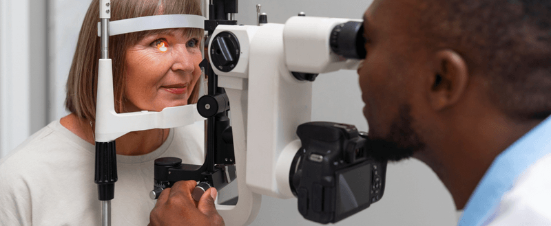
ICL surgery takes 20-30 minutes per eye and is performed under local anaesthetic.  A small opening is made in the periphery of the cornea to allow the ICL to be introduced into the eye through an injector. Once inside the eye, the lens unfolds and is positioned behind the iris. The small opening is self-sealing and does not require any sutures.


Visual recovery is fast and most patients are able to see very well by the next day. Your vision will continue to improve as the eye heals and settles down over the first few weeks following ICL surgery

Results You Can Expect With an Implantable Collamer Lens

ICL is designed to deliver strong visual outcomes in appropriate candidates, but it is important to set realistic expectations and understand long-term factors.PresbyMAX is one of several ways to manage presbyopia. The best option depends on your eyes, your goals, and how you feel about different trade-offs.

Visual Outcomes and Stability

The goal is sharp, stable vision with reduced dependence on glasses or contact lenses. Some people still choose to use glasses for specific tasks, but many achieve meaningful day-to-day freedom from corrective eyewear.

Night Vision and Halos

Some people notice halos or glare, particularly in dim lighting. This is assessed before surgery by looking at pupil size, prescription, and your visual needs. If night driving is a major part of your life, that should be discussed during planning so expectations are clear.

Long-Term Considerations

Even with excellent distance vision after ICL, vision can change with age. Many people will still need reading glasses later due to normal age-related focusing changes. Cataracts can also develop later in life because that is part of ageing. If cataracts develop, the ICL can be removed at the time of cataract surgery if required.

Why Choose City Eye Surgeons Clinic for ICL

Choosing the right setting for ICL is about careful assessment and a safety-first approach, not just the procedure day.

Comprehensive Suitability Screening

ICL is only recommended when measurements and eye health checks support a safe outcome. Screening includes anatomical safety checks, pressure risk assessment, and a close look at the retina when myopia is higher.

Surgeon Experience and Safety-First Planning

Lens selection, sizing accuracy, and surgical technique all matter. Your surgeon will explain why a particular plan is being recommended, what the main risks are for your eyes specifically, and how those risks are managed.

Ongoing Aftercare and Long-Term Monitoring

Follow-up is a core part of ICL care. Monitoring focuses on eye pressure, lens position, and long-term eye health so any changes can be managed early.

Book a Consultation
If you’d like to find out more about implantable collamer lens, please call us on (03) 9070 0955, or contact us using the online form below and we will be in touch.

FAQs about Implantable Collamer Lens

Can ICL correct astigmatism?

The difference between ICL and LASIK is that ICL corrects vision by implanting a lens inside the eye while LASIK corrects vision by creating a corneal flap and reshaping corneal tissue with a laser.

Yes. Toric ICL options are designed to correct astigmatism, but careful measurement and alignment are important.

Do not drive on the day of surgery. You can drive again once your vision is safe and you have been cleared at follow-up. This timing varies between patients.

Implantable Collamer Lens (ICL)

Freedom from glasses starts here.

An implantable collamer lens, often called ICL, is a vision correction option for people who want to reduce their reliance on glasses or contact lenses without reshaping the cornea. Instead of using laser to change the front surface of the eye, an ICL adds a lens inside the eye to correct your prescription. This can be a strong option for higher prescriptions, thin corneas, or people who are concerned about dry eye with some laser procedures.

What is glaucoma

Glaucoma is diagnosed based on optic nerve changes and functional vision testing, not just a single eye pressure reading. Understanding how glaucoma affects vision helps explain why regular screening and ongoing monitoring are so important.

How glaucoma affects the optic nerve

Glaucoma damages the optic nerve fibres over time. Because the nerve fibres responsible for peripheral vision are often affected first, many people do not notice any change early on. Central vision can stay clear until the disease is more advanced, which is why regular testing is so important. Without treatment, the field of vision can gradually narrow.

Is glaucoma the same as high eye pressure

Not exactly. High eye pressure is called ocular hypertension, and some people with ocular hypertension never develop glaucoma. On the other hand, some people develop glaucoma even when their eye pressure is in the normal range. This is known as normal tension glaucoma. Glaucoma is diagnosed based on optic nerve damage and/or characteristic visual field changes, not on pressure alone.
Types of glaucoma we diagnose and treat
Glaucoma is not one single condition. The type matters because it affects urgency, testing focus, and treatment choices.

Primary open angle glaucoma

This is the most common form. The drainage angle is open, but the fluid does not drain efficiently enough, so pressure can build over time and slowly damage the optic nerve.

Angle closure glaucoma

Angle closure happens when the drainage angle becomes narrow or blocked, preventing fluid from leaving the eye.

  • Acute angle closure can cause a sudden spike in pressure and severe symptoms. This is an emergency.
  • Chronic angle closure develops more gradually and can be picked up during an eye examination before an attack occurs.

Secondary glaucoma

Secondary glaucoma is caused by another factor affecting drainage or pressure. Examples include inflammation inside the eye, previous trauma, pigment dispersion, and a steroid response (from steroid drops, tablets, or inhalers).

Glaucoma symptoms and warning signs

Many people expect glaucoma to cause pain or noticeable blur early, but that is not usually the case. Knowing the difference between slow, symptom-free glaucoma and an acute attack is important.

Early glaucoma symptoms

In many cases, there are no symptoms at the start. Any changes tend to be subtle and may include gradual loss of peripheral vision that is easy to miss day to day.

Symptoms of acute angle closure

Acute angle closure can cause:

  • Sudden eye pain and redness
  • Blurred vision and halos around lights
  • Headache, nausea, or vomiting

If these symptoms occur, seek urgent care immediately.

Glaucoma risk factors

Who is more likely to develop glaucoma

Risk is higher if you have one or more of the following:

  • Increasing age
  • Family history of glaucoma
  • Higher eye pressure
  • Thinner corneas
  • Certain ethnic backgrounds (risk can vary by glaucoma type)
  • Higher levels of short-sightedness
  • Diabetes or vascular factors in some patients

If you have diabetes, it is also worth understanding retina-related risks such as diabetic retinopathy, which can affect vision in different ways.

Medications and conditions that can increase eye pressure

Steroids are a common trigger for raised pressure in susceptible people, including steroid eye drops, tablets, injections, creams used around the eyes, and some inhalers. Previous eye injury, inflammation, and certain eye conditions can also increase risk.

Glaucoma testing and diagnosis

Visual field testing

Visual field testing checks how well you see in your peripheral vision. It is one of the most important tools for monitoring glaucoma because it measures functional vision, not just structure.

OCT and imaging for glaucoma

OCT imaging can measure the retinal nerve fibre layer and ganglion cell complex. This can help detect early glaucoma changes and confirm whether treatment is keeping things stable.

Angle assessment and corneal thickness

Gonioscopy checks whether the drainage angle is open or narrow, which guides diagnosis and treatment decisions. Pachymetry measures corneal thickness because it can influence pressure readings and help inform a realistic target pressure.

Glaucoma diagnosis is built from several pieces of information collected over time. The goal is to confirm whether glaucoma is present, establish a baseline, and then track for change.

Eye pressure checks

Eye pressure is measured with tonometry. One reading is not enough to diagnose glaucoma because pressure can vary throughout the day and from visit to visit. Trends over time matter.

Optic nerve assessment

Your specialist will examine the optic nerve closely and may use imaging to document its shape and nerve fibre appearance. This helps detect early damage and track changes over time.

Glaucoma treatment options

Treatment focuses on lowering eye pressure to a level that is safer for the optic nerve. The best option depends on glaucoma type, severity, and how stable the optic nerve and visual fields are over time.

Glaucoma eye drops

Eye drops are commonly used to lower eye pressure by reducing fluid production or improving drainage. Consistent daily use matters. Side effects vary depending on the drop type and can include stinging, redness, or dry eye symptoms, so it is important to tell your doctor if anything feels off.

Laser treatment for glaucoma

Laser options may be used to lower pressure or reduce the need for drops.

  • SLT (Selective Laser Trabeculoplasty) is commonly used for open angle glaucoma to improve drainage.
  • Laser iridotomy can be used if the angle is narrow or at risk of closure.

Minimally invasive glaucoma surgery (MIGS)

MIGS procedures are designed to lower eye pressure with a smaller surgical footprint than traditional glaucoma surgery. They are often considered at the time of cataract surgery in suitable patients, depending on glaucoma type and target pressure needs.

Glaucoma surgery

When drops and laser are not enough, surgery may be recommended. This can include trabeculectomy or drainage implants (tubes). These procedures aim to create a new pathway for fluid to leave the eye and protect the optic nerve from further damage.

What to expect from glaucoma treatment

Treatment depends on your prescription, symptoms, corneal shape, and lifestyle goals. Some people want the sharpest possible correction for work and driving, while others are mainly trying to reduce the hassle of glasses or contacts.aGlaucoma care is a long-term plan, not a one-time fix. The focus is stability, protecting the optic nerve, and catching progression early if it occurs.

Setting a target eye pressure

A “safe” pressure is different for each person. Your target is based on optic nerve health, visual field results, rate of change (if any), and individual risk factors. The goal is stability over time.

Ongoing monitoring and follow-up

Glaucoma care is long-term. Follow-up usually includes repeat pressure checks, optic nerve review, OCT imaging, and visual field testing at intervals based on your level of risk and stability. Regular reviews help catch change early.

Can glaucoma be cured

There is no cure, and vision that has been lost cannot be restored. Treatment is focused on preserving the vision you have and reducing the risk of further loss.

Living with glaucoma

Most people with glaucoma live full lives, but success depends on daily habits and ongoing reviews. Small practical steps can make treatment easier and more effective.

Using eye drops correctly

A few practical habits can make drops easier and more effective:

  • Use the drop at the same time each day
  • If you use more than one drop, leave a gap of a few minutes between them
  • Try gentle eyelid closure after instilling drops to reduce runoff
  • If you miss doses often, tell your doctor so the plan can be simplified where possible

Driving and vision

Glaucoma can affect peripheral vision, which matters for driving safety. If you notice changes, or if you are unsure about your vision standards, discuss it early so you can get clear advice and appropriate testing.

Protecting your long-term eye health

Keep your appointments, stick with your treatment plan, and report side effects rather than pushing through them. Glaucoma is often manageable, but it relies on consistent follow-up.

Results you can expect with an implantable collamer lens

ICL is designed to deliver strong visual outcomes in appropriate candidates, but it is important to set realistic expectations and understand long-term factors.

Visual outcomes and stability

The goal is sharp, stable vision with reduced dependence on glasses or contact lenses. Some people still choose to use glasses for specific tasks, but many achieve meaningful day-to-day freedom from corrective eyewear.

Night vision and halos

Some people notice halos or glare, particularly in dim lighting. If night driving is a major part of your life, that should be discussed during planning so expectations are clear.

Long-term considerations

Even with excellent distance vision after ICL, vision can change with age. Many people will still need reading glasses later due to normal age-related focusing changes. Cataracts can also develop later in life because that is part of ageing. If cataracts develop, the ICL can be removed at the time of cataract surgery if required.

Why choose City Eye Surgeons for ICL surgery

Choosing the right setting for ICL is about careful assessment and a safety-first approach, not just the procedure day.

Ongoing aftercare and long-term monitoring

Follow-up is a core part of ICL care. Monitoring focuses on eye pressure, lens position, and long-term eye health so any changes can be managed early.

Comprehensive suitability screening

ICL is only recommended when measurements and eye health checks support a safe outcome. Screening includes anatomical safety checks, pressure risk assessment, and a close look at the retina when myopia is higher.

Surgeon experience and safety-first planning

Lens selection, sizing accuracy, and surgical technique all matter. Your surgeon will explain why a particular plan is being recommended, what the main risks are for your eyes specifically, and how those risks are managed.

FAQs

What is the difference between ICL and LASIK?
The difference between ICL and LASIK is that ICL corrects vision by implanting a lens inside the eye while LASIK corrects vision by creating a corneal flap and reshaping corneal tissue with a laser.
Yes. Toric ICL options are designed to correct astigmatism, but careful measurement and alignment are important.
The surgical portion is usually short, but you should plan for a longer total time in hospital for preparation, checks, and monitoring before you go home.
Do not drive on the day of surgery. You can drive again once your vision is safe and you have been cleared at follow-up. This timing varies between patients.

Book an ICL assessment in Melbourne

To book an assessment, contact the clinic and request an ICL suitability consultation. It helps to bring:

  • Your most recent glasses prescription
  • Your contact lens details, including brand and wearing schedule
  • Any previous glasses prescriptions if available
  • A list of current medications

Rediscover clearer vision